The Axial Skeleton Exercise 8 embarks on an exploration of the human skeletal system, delving into the intricate structure and functions of the axial skeleton, the foundational pillar of our bodies.
This exercise provides a comprehensive understanding of the axial skeleton’s components, organization, and clinical significance, shedding light on its vital role in movement, posture, and overall well-being.
Introduction to the Axial Skeleton
The axial skeleton is the central, vertical axis of the body that provides support, protection, and movement. It comprises the bones of the head, neck, and trunk.
The axial skeleton serves several essential functions:
- Protection:Encases and safeguards vital organs such as the brain, spinal cord, and heart.
- Support:Provides a rigid framework for the body, allowing it to maintain an upright posture.
- Movement:Enables a wide range of movements, including head rotation, neck flexion, and trunk bending.
Structure and Organization of the Axial Skeleton
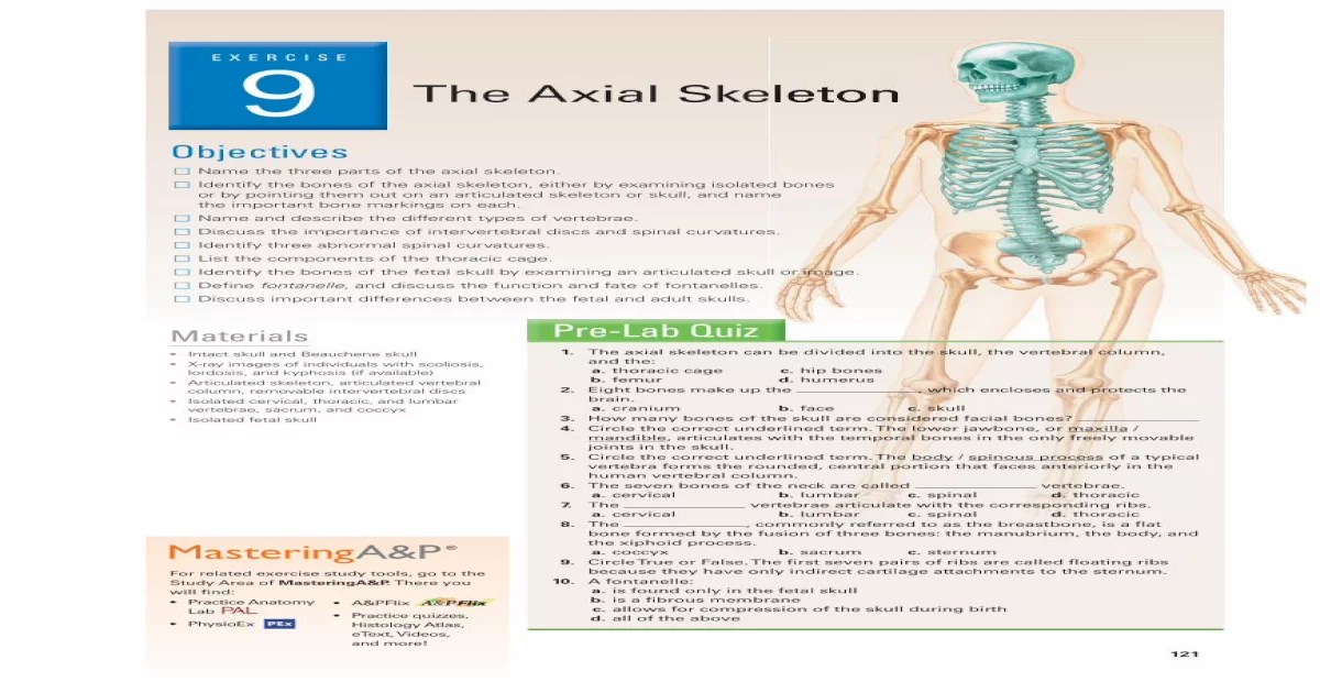
The axial skeleton forms the central axis of the body, providing support and protection for vital organs. It comprises the skull, vertebral column, and rib cage.The vertebral column, also known as the spine, consists of 33 vertebrae stacked one upon another.
Exercise 8 of the axial skeleton series delves into the intricacies of bone structure and articulation. For a comprehensive understanding of the axial skeleton, it’s recommended to supplement this exercise with a visual aid such as the court of the gentiles diagram . This diagram provides a detailed illustration of the axial skeleton, including the vertebrae, ribs, and sternum, offering a valuable perspective to complement the theoretical knowledge gained from the exercise.
Each vertebra has a central canal that houses the spinal cord. Between adjacent vertebrae lie intervertebral discs, which act as shock absorbers and allow for flexibility.
Rib Cage
The rib cage protects the thoracic cavity and its organs. It consists of 12 pairs of ribs, which attach posteriorly to the vertebrae and anteriorly to the sternum via costal cartilages. The sternum is a flat bone located in the midline of the chest.
Skull
The skull encloses and protects the brain. It comprises two main parts: the cranium and the facial bones. The cranium consists of eight bones that form the protective vault around the brain. The facial bones consist of 14 bones that form the face and provide openings for the eyes, nose, and mouth.
The nasal cavity is a space within the skull that houses the olfactory organs.
Muscles of the Axial Skeleton
The axial skeleton, comprising the skull, vertebral column, and rib cage, provides structural support and protection for vital organs. Various muscles attach to this framework, enabling movement, maintaining posture, and facilitating respiration.
Muscles of the Head
Numerous muscles are associated with the skull, contributing to facial expressions, chewing, swallowing, and head movement. Key muscles include:
- Temporalis:Elevates the mandible during chewing.
- Masseter:Closes the mandible, aiding in biting and grinding.
- Orbicularis Oculi:Closes the eyelids, protecting the eyes.
- Sternocleidomastoid:Rotates and laterally flexes the head.
Muscles of the Neck
The neck region houses muscles responsible for head and neck movements, including:
- Platysma:Tightens the skin of the neck.
- Scalenes:Flex the neck laterally and elevate the first two ribs during inspiration.
- Trapezius:Elevates and retracts the scapulae.
- Longus Colli:Flexes the neck anteriorly.
Muscles of the Back
The back muscles play a crucial role in supporting the vertebral column, maintaining posture, and facilitating trunk movements:
- Erector Spinae:Extends the vertebral column and assists in maintaining an upright posture.
- Multifidus:Rotates and laterally flexes the vertebral column.
- Transversus Abdominis:Compresses the abdomen and aids in exhalation.
- Rhomboids:Retract and elevate the scapulae.
Muscles of the Chest
The chest muscles are involved in respiration and arm movements:
- Pectoralis Major:Adducts and medially rotates the arm.
- Pectoralis Minor:Depresses the shoulder and stabilizes the scapula.
- Intercostal Muscles:Aid in inspiration and expiration by elevating and depressing the ribs.
- Diaphragm:Separates the thoracic and abdominal cavities, playing a vital role in respiration.
Joints of the Axial Skeleton: The Axial Skeleton Exercise 8
The axial skeleton, which comprises the skull, vertebral column, and rib cage, is held together by various types of joints. These joints allow for movement, support, and protection of the delicate structures within the axial skeleton.
The different types of joints found in the axial skeleton include:
Fibrous Joints
- Sutures:Immovable joints that connect the bones of the skull.
- Syndesmoses:Slightly movable joints that connect bones using ligaments, such as the joints between the vertebrae.
Cartilaginous Joints
- Synchondroses:Immovable joints that connect bones using cartilage, such as the growth plates in long bones.
- Symphyses:Slightly movable joints that connect bones using fibrocartilage, such as the joint between the pubic bones.
Synovial Joints, The axial skeleton exercise 8
- Plane Joints:Allow for gliding movements, such as the joints between the vertebrae.
- Hinge Joints:Allow for flexion and extension movements, such as the joints between the vertebrae and the ribs.
- Pivot Joints:Allow for rotation movements, such as the joint between the first and second cervical vertebrae (atlas and axis).
- Condyloid Joints:Allow for flexion, extension, abduction, and adduction movements, such as the joint between the mandible and the temporal bone.
- Saddle Joints:Allow for flexion, extension, abduction, adduction, and circumduction movements, such as the joint between the thumb and the carpal bones.
- Ball-and-Socket Joints:Allow for a wide range of movements, including flexion, extension, abduction, adduction, rotation, and circumduction, such as the joint between the humerus and the scapula.
Each type of joint has a specific structure and function that allows for a particular range of motion and stability. The fibrous joints provide stability and support, while the cartilaginous joints allow for some movement and flexibility. The synovial joints offer the greatest range of motion and are found in areas where flexibility and movement are essential.
Clinical Significance of the Axial Skeleton

The axial skeleton is prone to various injuries and disorders that can affect its structure and function. Understanding these conditions is crucial for proper diagnosis and treatment.
Common injuries include fractures, dislocations, and sprains, which can occur due to trauma, falls, or accidents. Disorders such as osteoporosis, arthritis, and scoliosis can also affect the axial skeleton, leading to pain, deformity, and impaired mobility.
Diagnostic and Treatment Options
Diagnosis of axial skeleton injuries and disorders involves a combination of physical examination, imaging techniques (X-rays, CT scans, MRI), and medical history.
Treatment options vary depending on the specific condition. Fractures and dislocations may require immobilization, casting, or surgery. Osteoporosis can be managed with medication, lifestyle modifications, and calcium and vitamin D supplements. Arthritis can be treated with pain relievers, anti-inflammatory drugs, and physical therapy.
Importance of Proper Posture and Spinal Health
Maintaining proper posture is essential for spinal health. Poor posture can lead to muscle strain, back pain, and spinal deformities. Regular exercise, ergonomic workstations, and proper sleeping positions can help maintain good posture.
Spinal health involves preserving the natural curvature of the spine. Deviations from the normal curvature, such as scoliosis, kyphosis, and lordosis, can cause pain, discomfort, and impaired mobility. Regular exercise, physical therapy, and, in severe cases, surgery can help correct spinal deformities.
Quick FAQs
What is the axial skeleton?
The axial skeleton comprises the bones of the head, neck, and trunk, forming the central axis of the body.
What are the main functions of the axial skeleton?
The axial skeleton provides structural support, protects vital organs, facilitates movement, and houses the central nervous system.
What are some common injuries and disorders of the axial skeleton?
Common injuries include fractures, sprains, and dislocations, while disorders include osteoporosis, arthritis, and spinal cord injuries.